English
 norsk
norskBlar i forfatter "Lindgren, Peter"
Viser treff 1-6 av 6
-
Automated analysis of color tissue Doppler velocity recordings of the fetal myocardium using a new algorithm
(Journal article; Tidsskriftartikkel; Peer reviewed, 2015-08-27)Background: Tissue Doppler imaging (TDI) can be used to assess fetal cardiac function and it has been shown to detect changes associated with hypoxia in animal models. However, the analysis is cumbersome and time consuming. The main objective of this study was to evaluate the feasibility of a new algorithm developed for the automated analysis of color TDI velocity recordings of the fetal myocardium. ... -
Automated analysis of fetal cardiac function using color tissue Doppler imaging in second half of normal pregnancy
(Journal article; Tidsskriftartikkel, 2018-02-26)<i>Objectives</i> - Color tissue Doppler imaging (cTDI) is a promising tool for the assessment of fetal cardiac function. However, the analysis of myocardial velocity traces is cumbersome and time‐consuming, limiting its application in clinical practice. The aim of this study was to evaluate fetal cardiac function during the second half of pregnancy and to develop reference ranges using an ... -
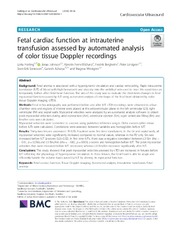
Fetal cardiac function at intrauterine transfusion assessed by automated analysis of color tissue Doppler recordings
(Journal article; Tidsskriftartikkel; Peer reviewed, 2020-08-13)Background: Fetal anemia is associated with a hyperdynamic circulation and cardiac remodeling. Rapid intrauterine transfusion (IUT) of blood with high hematocrit and viscosity into the umbilical vein used to treat this condition can temporarily further affect fetal heart function. The aim of this study was to evaluate the short-term changes in fetal myocardial function caused by IUT using automated ... -
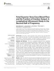
Fetal Superior Vena Cava Blood Flow and Its Fraction of Cardiac Output: A Longitudinal Ultrasound Study in the Second Half of Pregnancy
(Journal article; Tidsskriftartikkel; Peer reviewed, 2021-07-06)<i>Introduction</i>: In the fetus, a large proportion of the superior vena cava blood flow (QSVC) comes from the brain. To provide the possibility of using this blood flow as a representation of fetal brain circulation, we aimed to determine the fetal QSVC and its fraction of cardiac output during the second half of physiological pregnancies.<br><br> <i>Materials and Methods</i>: This was a ... -
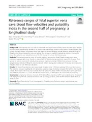
Reference ranges of fetal superior vena cava blood flow velocities and pulsatility index in the second half of pregnancy: a longitudinal study
(Journal article; Tidsskriftartikkel; Peer reviewed, 2021-02-23)Background - Fetal superior vena cava (SVC) is essentially the single vessel returning blood from the upper body to the heart. With approximately 80-85% of SVC blood flow representing cerebral venous return, its interrogation may provide clinically relevant information about fetal brain circulation. However, normal reference values for fetal SVC Doppler velocities and pulsatility index are lacking. ... -
Volume blood flow-based indices of fetal brain sparing in the second half of pregnancy: A longitudinal study
(Journal article; Tidsskriftartikkel; Peer reviewed, 2020-07-07)<i>Introduction</i> - Cerebroplacental ratio (CPR) and umbilicocerebral ratio (UCR) are clinically used as a measure of fetal brain sparing. These are calculated as the ratios between the pulsatility indices (PIs) of middle cerebral (MCA) and umbilical (UA) arteries, and are an indirect representation of the balance between cerebral and placental perfusion. Volume blood flow (Q)‐based ratios, ie ...